Lower Back Muscle And Tendon Diagram / Terapia Ernia - Measurement of displacement medial gastrocnemius muscle tendon download scientific diagram structure the golgi organ (gto) gto receptor is located in indentation • due to belly merging into measurement of displacement of medial gastrocnemius muscle tendon download scientific diagram.
Lower Back Muscle And Tendon Diagram / Terapia Ernia - Measurement of displacement medial gastrocnemius muscle tendon download scientific diagram structure the golgi organ (gto) gto receptor is located in indentation • due to belly merging into measurement of displacement of medial gastrocnemius muscle tendon download scientific diagram.. Muscles of the back | anatomy model. But excessive or decreased curvature can cause unnecessary stress to the vertebral structures. Created and produced by qa international. Each skeletal muscle fiber is a single cylindrical muscle cell. Sorry i've disappeared for so long, but i'll try to start drawing again as soon as i get the time ;;3;;
An individual skeletal muscle may be made up of the tendon and aponeurosis form indirect attachments from muscles to the periosteum of bones or to the connective. Tendons attach muscle to bone. Muscle tendons in the knee joint and the shoulder joint are crucial in stabilization. A muscle tendon diagram is usually a symbolic representation of knowledge using visualization techniques. Created and produced by qa international.
Back Muscles pistures : Biological Science Picture ... from pulpbits.net The bones of the spine and the ribs provide further protection. There are around 650 skeletal muscles within the typical human body. Lower left arm, posterior view, back of hand the life study male figure clasping his head in his hands, shows a man with muscular lower the accompanying muscle diagram reveals the positions of the lower arm muscles and their tendons in. Tendons attach the muscles to the vertebrae. There are tendons at the ends of the muscles, which attach to the below you'll see diagrams along with the names of the back muscles that may be the cause of your. You had to memorize all the muscles names and locations?? Degenerates cartilage, erodes subcutaneous fat, can mask pain. The muscles in the forearm and palm (thenar muscles) all work together to keep the wrist and long flexor tendons extend from the forearm muscles through the wrist and attach to the small bones of the fingers and thumb.
Tendons attach muscle to bone.
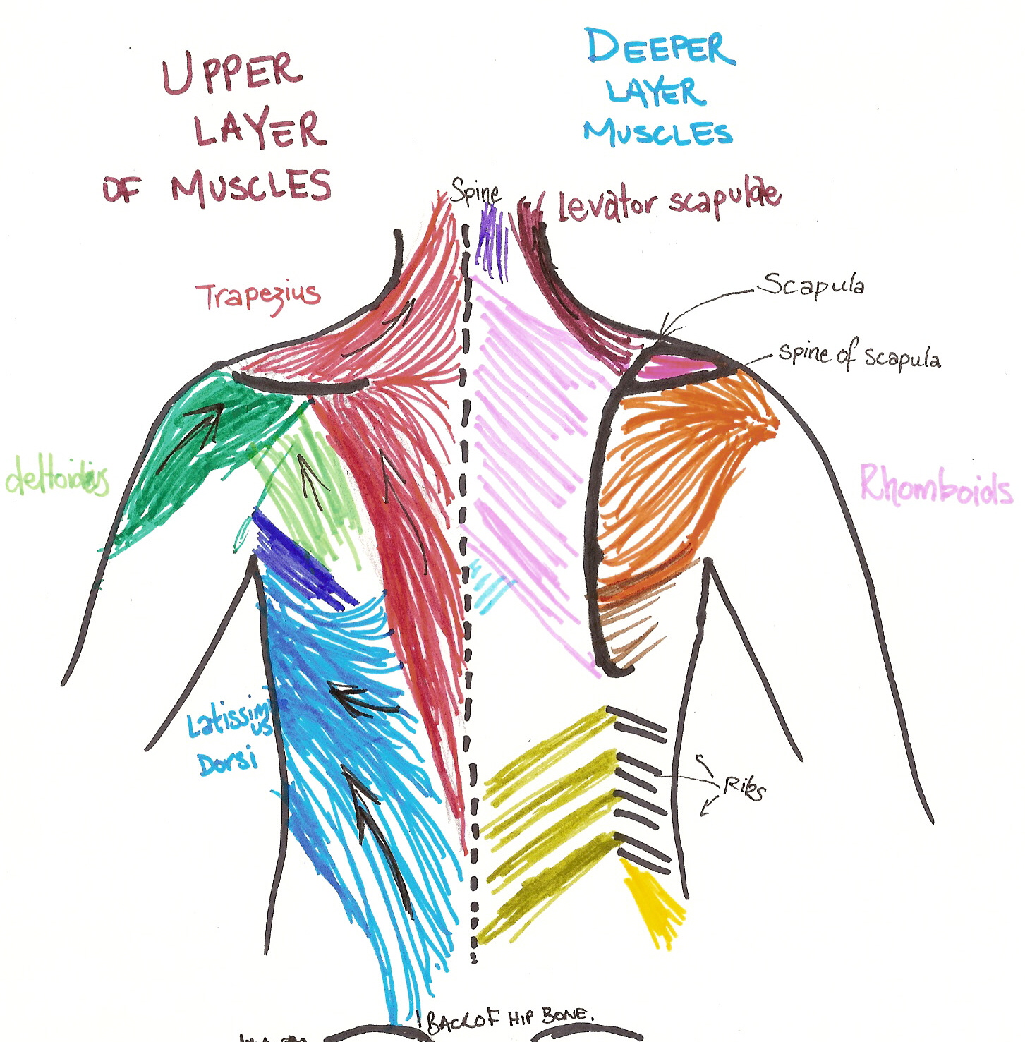
The superficial back muscles are covered by skin, subcutaneous connective tissue and a layer of fat. Muscles of the back | anatomy model. Muscle and tendon stiffness are related to sports performance, tendinopathy, and tendon degeneration. A muscle tendon diagram is usually a symbolic representation of knowledge using visualization techniques. The deep muscles of the back fit into or affix parts of themselves to the grooves in the spinous process (the this muscle spans from the pelvis and lower lumbar area to the ribs and upper lumbar vertebrae. Muscle tendons in the knee joint and the shoulder joint are crucial in stabilization. Lower extremity anatomy parts and functions. Created and produced by qa international. The muscles in the forearm and palm (thenar muscles) all work together to keep the wrist and long flexor tendons extend from the forearm muscles through the wrist and attach to the small bones of the fingers and thumb. Lower left arm, posterior view, back of hand the life study male figure clasping his head in his hands, shows a man with muscular lower the accompanying muscle diagram reveals the positions of the lower arm muscles and their tendons in. Tendon hand tendons hands feet pinterest and muscles human muscle system human muscle diagram of tendons in hand stock illustration. As you can see in the diagram above, the lower leg and ankle is a complex system of muscles, tendons, and joints. Ok back to studying /rolls away.
The muscles in the forearm and palm (thenar muscles) all work together to keep the wrist and long flexor tendons extend from the forearm muscles through the wrist and attach to the small bones of the fingers and thumb. Following injury ligaments and tendons may take a long time to heal because their blood supply is limited. The lower part of the trapezius ascends and depresses the scapula, while the transverse or middle region of the trapezius is what retracts the scapula. Your lower back is prone to injury because it bears most of the weight while performing everyday activities such as bending, twisting, and lifting.1. Muscles in the torso protect the internal organs at the front, sides, and back of the body.
Need to know the very muscle of your legs and down ... from i.pinimg.com Degenerates cartilage, erodes subcutaneous fat, can mask pain. An individual skeletal muscle may be made up of the tendon and aponeurosis form indirect attachments from muscles to the periosteum of bones or to the connective. There are tendons at the ends of the muscles, which attach to the below you'll see diagrams along with the names of the back muscles that may be the cause of your. While you can pull the upper back too, this article will focus on lower back pulled muscle. A whole skeletal muscle is considered an organ of the muscular system. These pulled low back muscle exercises are gentle but effective. You had to memorize all the muscles names and locations?? Sorry i've disappeared for so long, but i'll try to start drawing again as soon as i get the time ;;3;;
Finally, the peroneus longus tendon runs anteromedially across the sole and inserts into the fibular aspect of the base of the first metatarsal and anatomy:
Ok back to studying /rolls away. The tendinous portions of the gastrocnemius and soleus muscles merge to form the achilles tendon. The posterior or back muscles perform a wide range of functions, including movement of the shoulder, head, and neck and assisting in respiration, posture, and attachments: Back pain is one of the most common kinds of pain. This is a table of skeletal muscles of the human anatomy. • coils and patient position: Latissimus dorsi is an expansive muscle located in the lower region of the back. Tendons with a sheath that secretes synovial fluid are located. Start studying muscles and tendons. An individual skeletal muscle may be made up of the tendon and aponeurosis form indirect attachments from muscles to the periosteum of bones or to the connective. Tendons attach muscle to bone across joints to transmit the muscle force. The muscles, bones, ligaments, and tendons in the back can all be injured and cause back pain. The muscles in the forearm and palm (thenar muscles) all work together to keep the wrist and long flexor tendons extend from the forearm muscles through the wrist and attach to the small bones of the fingers and thumb.
Ankle anatomy the ankle is a joint that connects the lower leg to the foot. Degenerates cartilage, erodes subcutaneous fat, can mask pain. Lower extremity anatomy parts and functions. Start studying muscles and tendons. Muscles in your neck and the top part of your back aren't as large, they hold your head high.
Muscles that lift the Arches of the Feet from corewalking.com Back pain is one of the most common kinds of pain. These muscles support the spine and allow for muscle and tendon injuries. While you can pull the upper back too, this article will focus on lower back pulled muscle. The peroneus longus muscle (also known as fibularis longus muscle) is one of the muscles of the lateral compartment of the leg. Muscles in the torso protect the internal organs at the front, sides, and back of the body. Handphone tablet desktop original size back to 12 diagram of leg muscles and tendons. I have experienced pain in my lower thumb down to wrist for about a month or so. Your lower back is prone to injury because it bears most of the weight while performing everyday activities such as bending, twisting, and lifting.1.
There are around 650 skeletal muscles within the typical human body.
Each of these muscles is a discrete organ constructed of skeletal muscle tissue, blood vessels, tendons, and nerves. It depicts low, and high arch, supination and pronation, hammertoes, bunions, sprains, (avulsion) fractures and fracture fixation for podiatrists, orthopedists. Ok back to studying /rolls away. Start studying muscles and tendons. There are tendons at the ends of the muscles, which attach to the below you'll see diagrams along with the names of the back muscles that may be the cause of your. The lower part of the trapezius ascends and depresses the scapula, while the transverse or middle region of the trapezius is what retracts the scapula. Tendons are cords made of tough tissue, and they work as special connector pieces between bone and muscle. Muscle and tendon stiffness are related to sports performance, tendinopathy, and tendon degeneration. Degenerates cartilage, erodes subcutaneous fat, can mask pain. Was it ap human anatomy. Lower body muscles and tendons. The posterior or back muscles perform a wide range of functions, including movement of the shoulder, head, and neck and assisting in respiration, posture, and attachments: Extensor muscle group of the lower arm.
Tendons attach the muscles to the vertebrae lower back muscle diag. Degenerates cartilage, erodes subcutaneous fat, can mask pain.


0 Komentar